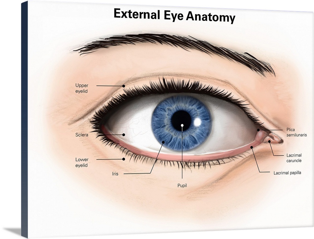
External anatomy of the human eye (with labels) Wall Art, Canvas Prints
Lacrimal gland Eye muscles Eyeball Outer layer Middle layer Inner layer Blood supply of the eye Nerves of the eye Sources + Show all Bones of the orbit The bony orbit is made out of seven bones, which include the maxilla, zygomatic bone, frontal bone, ethmoid bone, lacrimal bone, sphenoid bone and palatine bone.

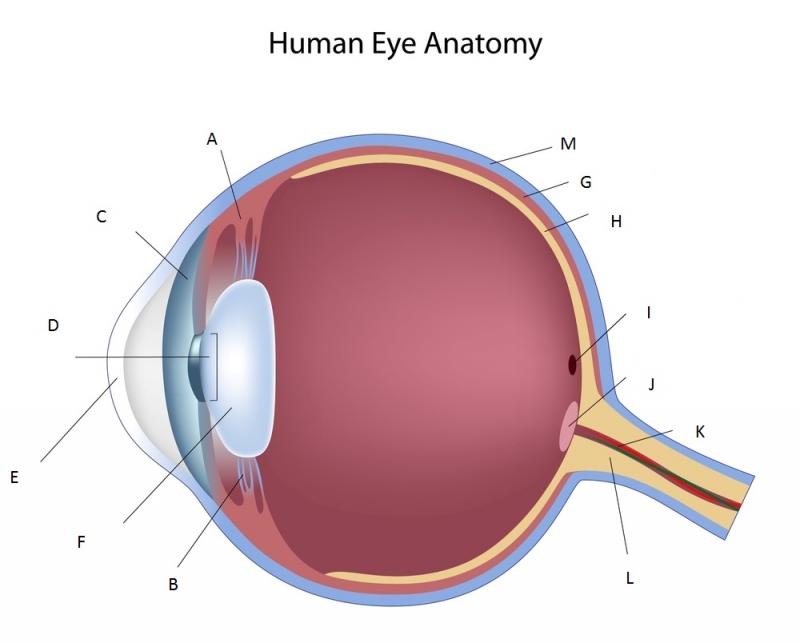
Diagram Of Human Eye Without Label
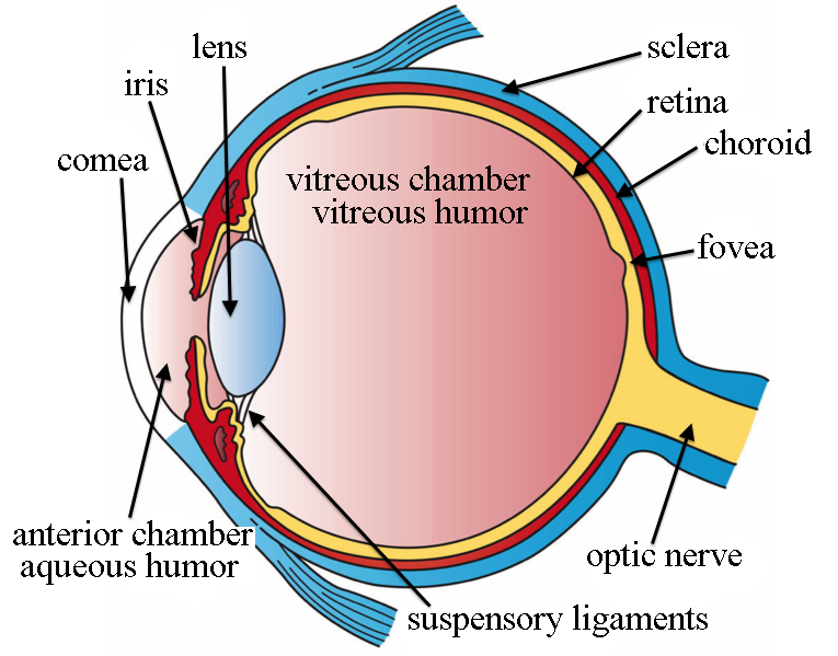
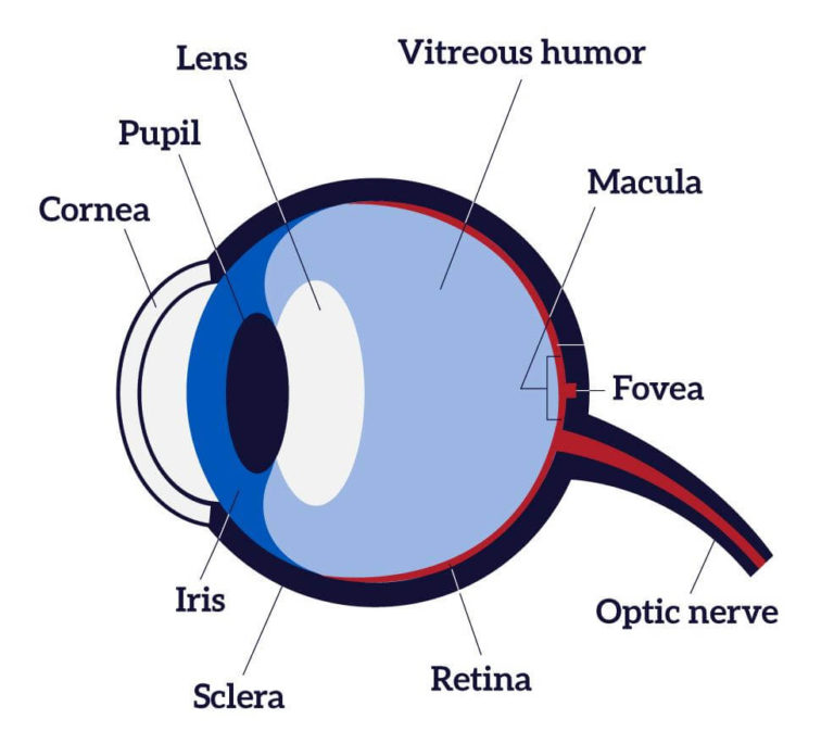
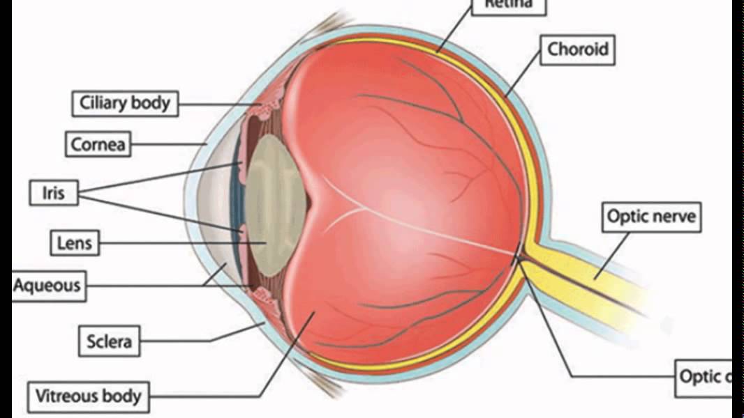
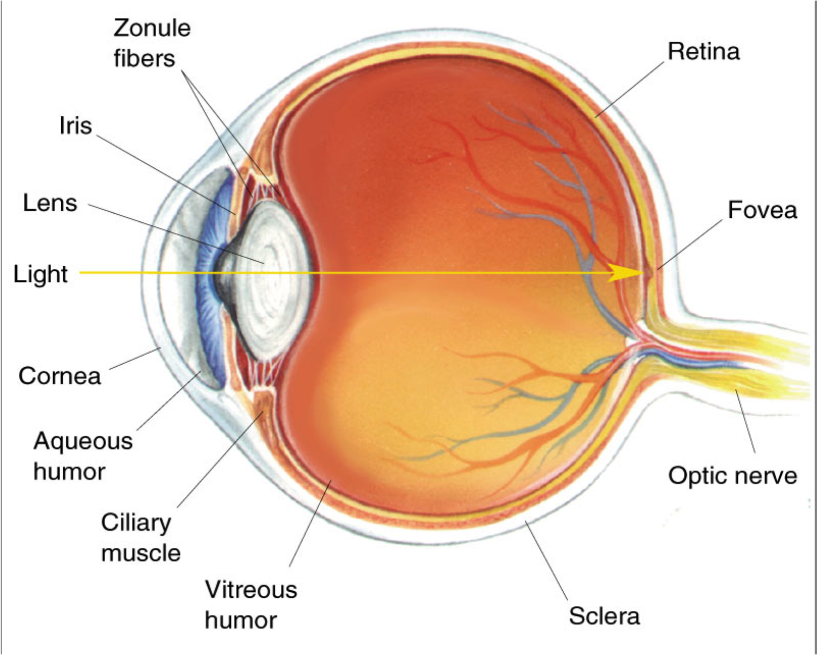
Eyes Anterior chamber. The front section of the eye's interior where aqueous humor flows in and out, providing nourishment to the eye. Aqueous humor. The clear watery fluid in the front of the eyeball. Blood vessels. Tubes (arteries and veins) that carry blood to and from the eye. Caruncle.

priseaden eye diagram labeled
Understanding how vision works When surveyed about the five senses — sight, hearing, taste, smell and touch — people consistently report that their eyesight is the mode of perception they value (and fear losing) most.
Labeled Parts Of The Eye ClipArt Best
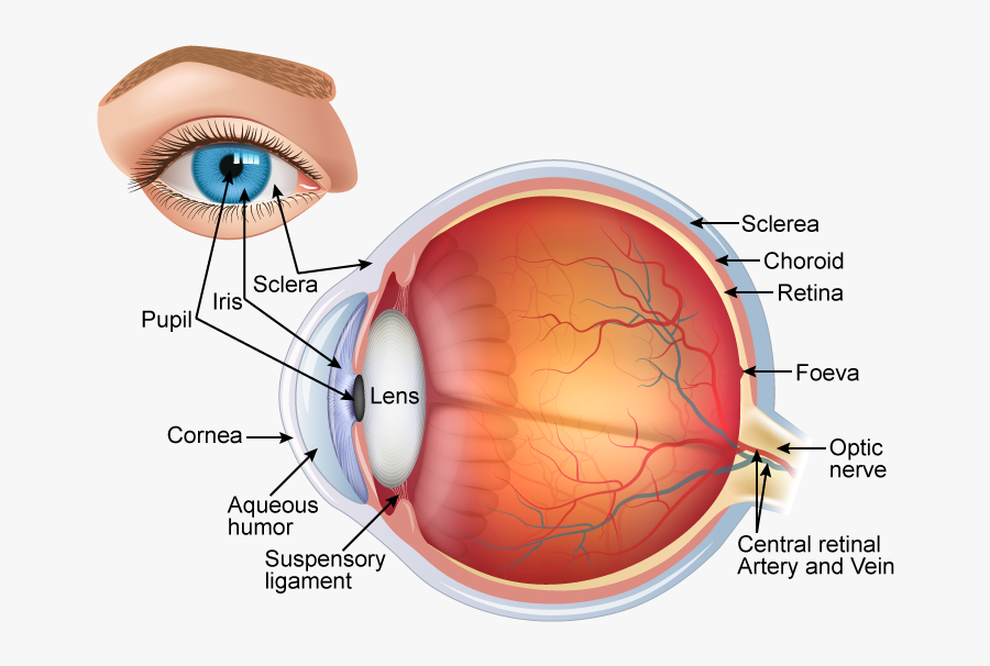
Contact Harvard Eye Associates at 949-951-2020 or harvardeye.com to schedule an appointment today. The eye is one of the most complicated organs in the human body. Major parts of the eye include the cornea, pupil, lens, retina and macula. Starting from "front" to "back" of the eye, the cornea is in charge of shaping the light as it.
Label The Eye ClipArt Best
Iris: regulates the amount of light that enters your eye. It forms the coloured, visible part of your eye in front of the lens. Light enters through a central opening called the pupil. Pupil: the circular opening in the centre of the iris through which light passes into the lens of the eye. The iris controls widening and narrowing (dilation and.
:max_bytes(150000):strip_icc()/GettyImages-695204442-b9320f82932c49bcac765167b95f4af6.jpg)
Structure and Function of the Human Eye
The process behind our vision: "The eye is a container," says Richard Rosen, M.D., a vitreoretinal surgeon at the New York Eye and Ear Infirmary of Mount Sinai, in New York City. The outside layer of the eye is a tough white protective layer called the sclera (more commonly known as the "white of the eye").
Human Eye Labelled Diagram , Free Transparent Clipart ClipartKey
1. Conjunctiva The conjunctiva is the membrane covering the sclera (white portion of your eye). The conjunctiva also covers the interior of your eyelids. Conjunctivitis, often known as pink eye, occurs when this thin membrane becomes inflamed or swollen. Other eye disorders that affect the conjunctiva include:
Anatomy of the eye Quizzes and diagrams Kenhub
Pupil: The pupil is the opening at the center of the iris. The iris adjusts the size of the pupil and controls the amount of light that can enter the eye. Retina: The retina is the light-sensitive tissue at the back of the eye. The retina converts light into electrical impulses that are sent to the brain through the optic nerve.
Human Eye Diagram, How The Eye Work 15 Amazing Facts of Eye
The structures and functions of the eyes are complex. Each eye constantly adjusts the amount of light it lets in, focuses on objects near and far, and produces continuous images that are instantly transmitted to the brain. The orbit is the bony cavity that contains the eyeball, muscles, nerves, and blood vessels, as well as the structures that.
35 Labeled Eye Diagram Wiring Diagram List
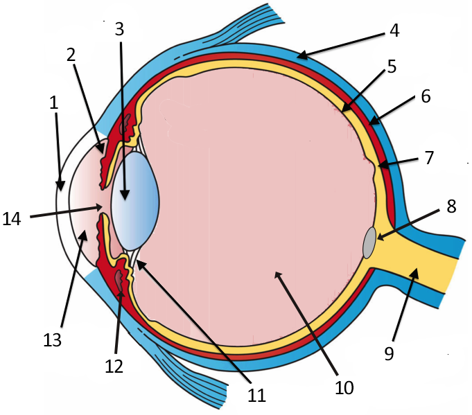
On a diagram of the eye, we can see all of the relevant structures together on one image. This helps us to understand how each one is situated and related to the other. Labeled diagram of the eye Diagram showing the parts of the eye with labels So, how can you use them to your benefit? Take a look at the diagram of the eyeball above.

Brain Post How Big is Your Blind Spot? Human eye
The optic foramen, the opening through which the optic nerve runs back into the brain and the large ophthalmic artery enters the orbit, is at the nasal side of the apex; the superior orbital fissure is a larger hole through which pass large veins and nerves.

33 Label Eye Labels 2021
Anatomy of the Human Eye. Eyes are one of the most important organs of the body. A healthy pair of eyes means a clear vision, which plays a major role in day-to-day life and quality of experiences.
Label the Eye
The eye has several major components: the cornea, pupil, lens, iris, retina, and sclera. These work together to capture an image and transmit it directly to the brain's occipital lobe via the.
Clip Art Details Eye Diagram Without Labels Free Transparent PNG
Apr. 29, 2023 To understand the diseases and conditions that can affect the eye, it helps to understand basic eye anatomy. Here is a tour of the eye starting from the outside, going in through the front and working to the back. Eye Anatomy: Parts of the Eye Outside the Eyeball The eye sits in a protective bony socket called the orbit.

Eye Model Labeled Bing Images Eye anatomy, Eye health, Eye sight
Glaucoma Material Type: Handouts Audience: Hispanics/Latinos Download English: Parts of the Eye (PDF 603.5 KB) Spanish: Las partes del ojo (PDF 897.7 KB) Check out this fact sheet to see a labeled diagram of the eye and learn about the different parts of the eye.
Eye Diagram Cliparts.co
Diabetes Healthy ANATOMY and Eyes OF THE AND ITS FUNCTION Toolkit Parts of the Eye Vision is wonderful, but you could lose To understand it if you eye have problems, diabetes. it is helpful to know the different parts of the eye. Please refer to the back of this handout for descriptions of their functions. The main parts of the eye— Optic 3